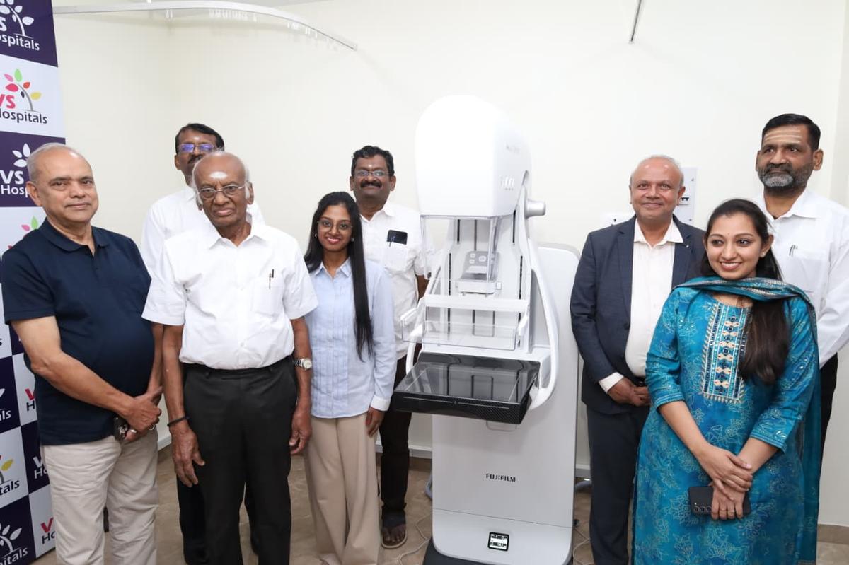
Early detection saves lives. Catching abnormal tissue before a lump can even be felt by hand remains the sharpest defense against breast cancer, and Chennai's VS Hospitals has upgraded its diagnostic arsenal to do just that.
The hospital launched Fujifilm's AMULET SOPHINITY 2D digital mammography system at its Chennai facility. Built around an ultra-fine 50-micron detector, the platform captures high-resolution breast tissue scans in seconds, revealing tiny specks of calcium known as microcalcifications—often the very first physical footprint of early-stage breast disease.
Early detection is essential in breast cancer care, and mammography plays a critical role in identifying abnormalities before symptoms emerge," said Dr. S. Subramanian, Chairman and Managing Director of VS Hospitals, during the unveiling.
Standard physical examinations routinely miss microscopic internal changes. Digital mammography solves this hurdle by passing low-dose X-rays through tissue to project high-contrast structural images onto digital sensors.
The newly installed Fujifilm unit sharpens that visibility.
By shrinking pixel size to 50 microns, the detector delivers clear diagnostic clarity. That fine resolution allows radiologists to spot minute structural distortions and suspicious microcalcifications that standard lower-resolution units can blur together.
Speed also matters when a patient is holding their breath in an exam room. The system clocks standard image acquisition in approximately five seconds. For detailed high-resolution imaging runs, the capture cycle finishes in 12 seconds flat.
| Technical Parameter | AMULET SOPHINITY Specification |
|---|---|
| Detector Pixel Resolution | 50 microns |
| Standard Image Acquisition Time | ~5 seconds |
| High-Resolution Capture Time | 12 seconds |
| Imaging Modality | 2D Digital Mammography (Low-Dose X-Ray) |
| Core Diagnostic Target | Microcalcifications & Non-palpable Lesions |
| Exposure Modulation | Automated Tissue Composition Matching |
Fear of physical pain keeps thousands of women from booking their annual screening. Traditional mammograms rely on heavy, rigid compression plates that clamp breast tissue flat, often causing acute discomfort.
The new setup directly targets that barrier.
Engineers equipped the unit with flexible positioning arms, intelligent breast compression mechanisms, and an automatic decompression release that eases pressure the moment exposure finishes. On-board artificial intelligence guides the radiographer, aligning the patient into position so scans come out right on the first try without grueling retakes.
Radiation dosage adjusts automatically as well. The system's intelligent exposure engine calculates tissue density in real time, dialing the X-ray beam up or down to ensure optimal contrast at the lowest possible radiation burden.
High-end medical hardware means little if patients cannot afford the diagnostic bill. To mark the installation, hospital administrators announced that VS Hospitals will run free mammography screenings for underprivileged individuals throughout the entire month of November 2026.
Patients and families seeking to register for the diagnostic initiative can reach hospital coordinators directly at 7448850500.