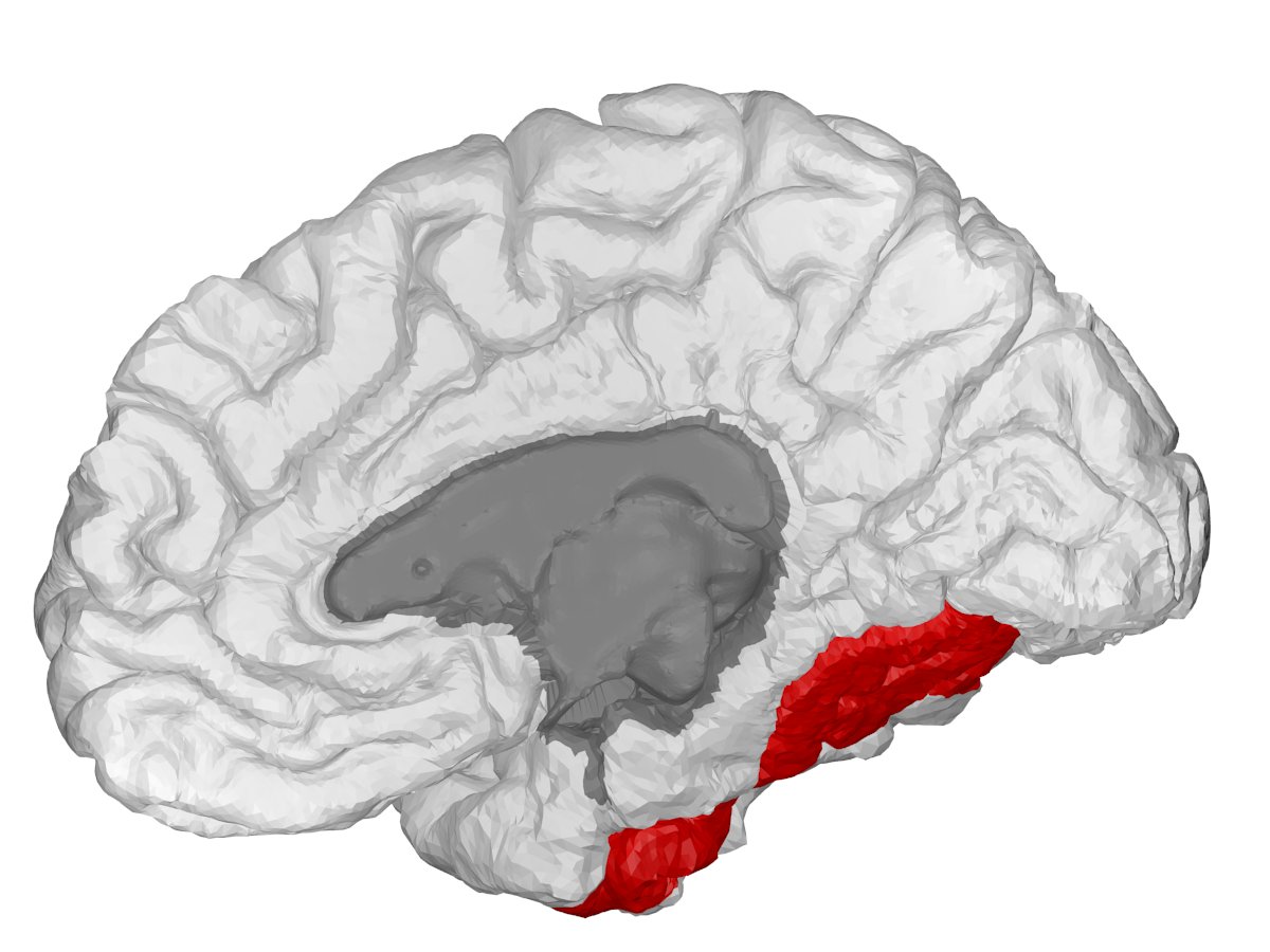
Scientists have cracked open the inner circuitry of the human basal ganglia. The breakthrough maps out 21 million cells across the deep brain command center that directs movement, habits, decision-making, and emotional balance. For decades, medicine treated this dense cluster of subcortical nuclei as an impenetrable black box. Now, investigators have built an atomic-grade blueprint tracking exactly which genes turn on, where specific cell types reside, and how molecular switches misfire during neurodegenerative collapse.
Basal ganglia malfunction drives severe neurological and psychiatric conditions, including Parkinson’s disease, Huntington’s disease, schizophrenia, Tourette’s syndrome, obsessive-compulsive disorder, and chemical addiction. The findings, detailed in more than two dozen studies published in Cell and affiliated journals, establish the first cross-species cellular catalog for an entire major brain structure.
Situated deep beneath the cerebral cortex, the basal ganglia coordinates action. It filters competing impulses. It lets you walk smoothly across a room, stop yourself from speaking out of turn, or lock in a daily workout routine. Three linked structures anchor this network: the striatum, the globus pallidus, and the substantia nigra.
When neurons in the substantia nigra perish, dopamine dries up and Parkinson's sets in. Yet clinicians have long lacked high-resolution data showing which specialized cells fail first.
"Until now, there was no reference describing each cell type in the human basal ganglia together with how it regulates its genes," said Joseph Ecker, a molecular biologist and geneticist at the Salk Institute in La Jolla, California, and one of the principal research leaders. "In the near term, the atlas is a reference resource for researchers. We are studying a Parkinson’s gene and can now see which cell types turn that gene on, and which DNA switches are active in those cells."
Over the longer term, the atlas can help identify cell-type-specific targets for gene therapy and gene editing. It also gives a baseline for comparing diseased tissue with healthy tissue.
Human living tissue cannot be dissected for routine laboratory screening. To bypass that barrier, researchers retrieved post-mortem brain tissue from deceased human donors, examining 21 million human cells alongside tissue samples from mice, macaque monkeys, and marmosets.
| Species Examined | Primary Structures Profiled | Molecular Datasets Captured | Target Application |
|---|---|---|---|
| Human (Homo sapiens) | Striatum, Globus Pallidus, Substantia Nigra | Gene activity, epigenome markers, spatial localization | Parkinson's, Schizophrenia, OCD, Bipolar |
| Macaque Monkey | Striatum, Subcortical nuclei | Cross-species gene expression alignment | Primate behavioral and motor modeling |
| Marmoset Monkey | Deep basal nuclei | Epigenetic switches and cellular conservation | Neurological drug response testing |
Cross-referencing four distinct species revealed which brain circuits evolved recently and which mechanisms remained untouched across millions of years. Salk Institute neuroscientist and biochemist Margarita Behrens emphasized that animal models remain foundational for therapeutic validation.
"Understanding the cellular composition of this brain structure across species will allow for comparative analyses of function as well as connectivity patterns and response to pharmacological interventions," Behrens stated.
Hongkui Zeng, director of brain science at the Allen Institute in Seattle, noted that the international team relies directly on comparative models to test drug efficacies and probe the neural loops governing human motor control.
By layering three dimensions of data, the research team constructed a living diagnostic index. The first layer catalogues transcriptomics—the specific active genes within each cell. The second charts the epigenome, the dense chemical tags and proteins that clamp onto DNA strands to silence or trigger cellular machinery. The third layer provides spatial coordinates, pinning each distinct cell type directly to its anatomical neighborhood.
The analysis uncovered a previously unknown brain cell type directly implicated in schizophrenia and bipolar disorder. It isolated distinct cell-specific genetic alterations linked to autism spectrum disorders, alongside gene expression changes that accelerate biological brain aging.
Neurologists caution that clinical cures will not arrive tomorrow. Precision medicines engineered from these cell maps face years of laboratory validation, non-human primate trials, and regulatory human screening.
Still, the breakthrough marks an operational turning point for the US National Institutes of Health BRAIN Initiative, first launched in 2014. Zeng confirmed that the consortium has set 2028 as the target date to deliver a complete, multimodal cellular atlas spanning the entire human brain.
| Laboratory Mouse | Whole basal network | High-throughput functional tracing | Preclinical genetic and circuit interventions |